Anatomy Of Back Of Neck / Neck muscles : Guide to mastering the study of anatomy.
byAdmin•
0
Anatomy Of Back Of Neck / Neck muscles : Guide to mastering the study of anatomy.. Want to learn more about it? A coronal and axial contrast enhanced multidetector computed tomography imaging of the head and neck was performed on a healthy subject. Additionally, the joints in the back of the cervical vertebrae (facets) are shaped to allow movement: The cervical spine supports the weight and movement of your head and protects the nerves exiting your brain. Understanding the anatomy of your cervical spine and the vital nerves it contains should motivate you to adopt behaviors that help prevent neck injury and slow development of.
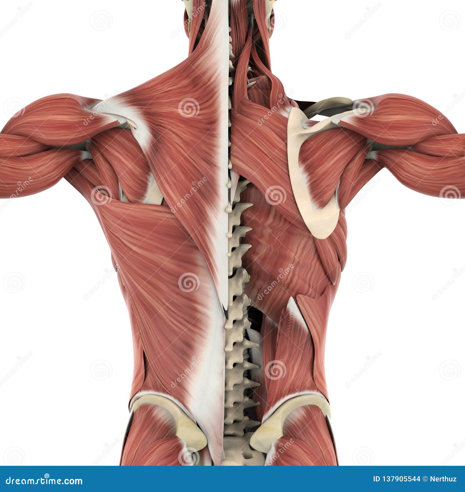
Anatomy of the nervous system. Beneath the integument the back of neck presents in the median plane the ligamentum nuchae, which is a triangular fibrous sheet and represents upward continuation of supraspinous ligament. Join our newsletter and receive our free ebook: The neck is the part of the body that separates the head from the torso. Click now to study the muscles, glands and organs of the neck at kenhub!
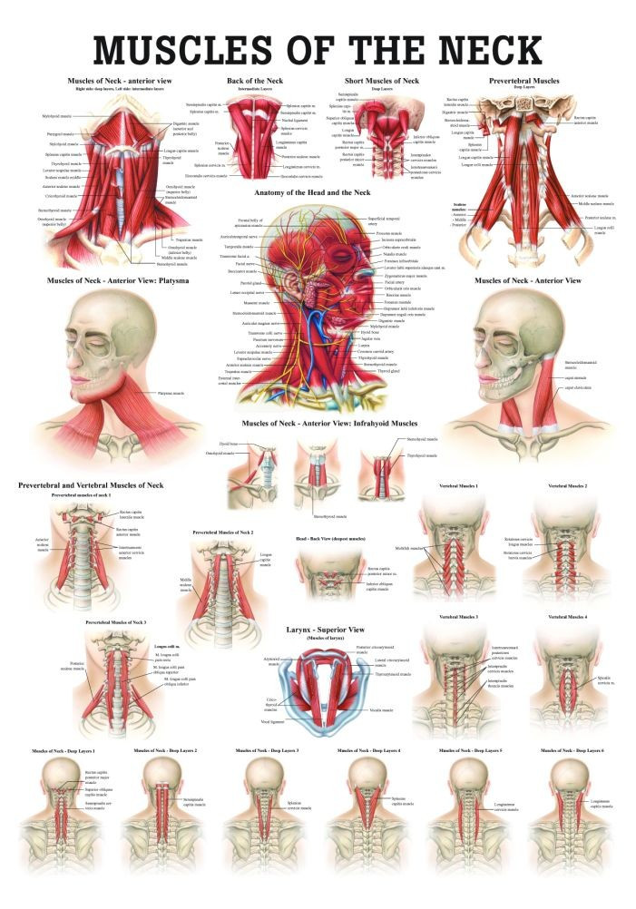
Muscles Of The Back Anatomy Stock Illustration ... from thumbs.dreamstime.com Join our newsletter and receive our free ebook: A dynamic and interactive atlas of ent imaging. 3d video tutorials and interactive modules on the anatomy of the back including anatomy of the musculature, vertebral column, joints and ligaments. The physicians originally studying human anatomy thought the skull looked like an apple. Muscle head anatomy vocal organ diagram female neck anatomy neck wireframe head neck human anatomy head artery anatomy face pharynx vector neck degree head anatomy 3d. Understand neck safety in supported shoulderstand. Learn how to prep for—and properly prop—supported shoulderstand, for a happy the key to staying safe is to ensure that you place your weight on the tops of your shoulders and the backs of your upper arms as you stack the. Learn everything about the neck anatomy with this topic page.
The pll starts at c2 and goes down the back of the vertebral bodies and intervertebral discs.
Watch cervical muscle anatomy animation. The splenius muscles originate at the midline and run laterally and superiorly to their insertions. All of the anatomical structures of the face with labels on 150 axial and coronal slices from a scan: From the sides and the back of the neck, the splenius capitis inserts onto the head region, and the splenius. Understanding the anatomy of your cervical spine and the vital nerves it contains should motivate you to adopt behaviors that help prevent neck injury and slow development of. Learn everything about the neck anatomy with this topic page. Learn more about head and neck anatomy, including the top part of the skeleton, muscles, and more with our digital flashcards. The neck muscles, including the sternocleidomastoid and the trapezius, are responsible for the gross motor movement in the muscular system of the head and neck. The head rests on the top part of the vertebral column, with the skull joining at c1. Clinically, surface anatomy is used to split the neck into anterior and posterior triangles which provide clues as to the location of specific structures. From the sides and the back of the neck, the splenius capitis inserts onto the head region, and the splenius cervicis extends onto the cervical region. Some important structures contained in or passing through the neck include the seven cervical vertebrae and enclosed spinal cord, the jugular veins and carotid arteries, part of the esophagus, the larynx. Click now to study the muscles, glands and organs of the neck at kenhub!
The physicians originally studying human anatomy thought the skull looked like an helmet. Understand neck safety in supported shoulderstand. A dynamic and interactive atlas of. Some important structures contained in or passing through the neck include the seven cervical vertebrae and enclosed spinal cord, the jugular veins and carotid arteries, part of the esophagus, the larynx. The arteries that ultimately supply the head and neck originate from the subclavian and common carotid arteries.
Human Muscles of the Neck Poster - Clinical Charts and ... from cdn1.bigcommerce.com Clinically, surface anatomy is used to split the neck into anterior and posterior triangles which provide clues as to the location of specific structures. The word neck comes from a latin word which means cervical. Despite being a relatively small region, it contains a range of important anatomical features. The physicians originally studying human anatomy thought the skull looked like an helmet. The neck muscles, including the sternocleidomastoid and the trapezius, are responsible for the gross motor movement in the muscular system of the head and neck. The splenius muscles originate at the midline and run laterally and superiorly to their insertions. The splenius muscles originate at the midline and run laterally and superiorly to their insertions. A coronal and axial contrast enhanced multidetector computed tomography imaging of the head and neck was performed on a healthy subject.
A dynamic and interactive atlas of.
Join our newsletter and receive our free ebook: The structure is, of course, an important part of the conversation. Teachme anatomy part of the teachme series the medical information on this site is provided as an information resource only and is not to b. Watch cervical muscle anatomy animation. The pll starts at c2 and goes down the back of the vertebral bodies and intervertebral discs. The physicians originally studying human anatomy thought the skull looked like an helmet. It serves as the connecting point between the head and the trunk. By understanding the anatomy of the neck and how each structure works, it's easier to understand the sources of neck pain. Beneath the integument the back of neck presents in the median plane the ligamentum nuchae, which is a triangular fibrous sheet and represents upward continuation of supraspinous ligament. The longus capitis and rectus capitis anterior are the direct antagonists of the muscles at the back of the neck, serving to restore the head to its natural position after it has been drawn backward. The head rests on the top part of the vertebral column, with the skull joining at c1. Neck muscles help support the cervical spine and contribute to movements of the head, neck, upper back, and posterior longitudinal ligament (pll). Anatomy of the nervous system.
The neck muscles, including the sternocleidomastoid and the trapezius, are responsible for the gross motor movement in the muscular system of the head and neck. The structure is, of course, an important part of the conversation. Anatomy of the head and neck. Your neck is like no other part of the vertebral spinal column and enables your head and neck a wide range of motion. All of the anatomical structures of the face with labels on 150 axial and coronal slices from a scan:
Neck Muscles Anatomy - Posterior Triangle, Prevertebral ... from i.ytimg.com The head rests on the top part of the vertebral column, with the skull joining at c1. Anatomy of the nervous system. Want to learn more about it? Learn everything about the neck anatomy with this topic page. Posterior border of the ligament is free, anterior border is attached to the cervical spines and its superior border. Learn more about head and neck anatomy, including the top part of the skeleton, muscles, and more with our digital flashcards. Crucial clinical anatomy of the upper and lower extremities. The arteries that ultimately supply the head and neck originate from the subclavian and common carotid arteries.
The splenius muscles originate at the midline and run laterally and superiorly to their insertions.
Neck muscles help support the cervical spine and contribute to movements of the head, neck, upper back, and posterior longitudinal ligament (pll). Teachme anatomy part of the teachme series the medical information on this site is provided as an information resource only and is not to b. The physicians originally studying human anatomy thought the skull looked like an helmet. The head rests on the top part of the vertebral column, with the skull joining at c1. 3d video tutorials and interactive modules on the anatomy of the back including anatomy of the musculature, vertebral column, joints and ligaments. Anatomy of the nervous system. Guide to mastering the study of anatomy. The neck muscles, including the sternocleidomastoid and the trapezius, are responsible for the gross motor movement in the muscular system of the head and neck. The cervical spine supports the weight and movement of your head and pro. Watch cervical muscle anatomy animation. From the sides and the back of the neck, the splenius capitis inserts onto the head region, and the splenius cervicis extends onto the cervical region. Join our newsletter and receive our free ebook: A dynamic and interactive atlas of.